Anatomy Of The Upper Chest Area : Muscles Of Upper Chest And Shoulder Stock Image C020 2123 Science Photo Library - The best upper chest workout will.
Dapatkan link
Facebook
X
Pinterest
Email
Aplikasi Lainnya
Anatomy Of The Upper Chest Area : Muscles Of Upper Chest And Shoulder Stock Image C020 2123 Science Photo Library - The best upper chest workout will.. Paschalides medical publications, 2004, with permission. Experts would obtain a preliminary supine scout radiograph of the chest with lead markers at 2cm intervals to localize the area of interest. The chest anatomy includes the pectoralis major, pectoralis minor and the serratus anterior. Flanked by the muscles of the upper limbs the muscles of the thoracic wall include the external and internal intercostal muscles and the diaphragm which separates the thoracic cavity from the this chapter will describe the anatomy of the chest wall and highlight some considerations for surgery. The cranial region encompasses the upper part of the head while the.
The upper posterior border of the heart is formed by the left atrium. • acromion • clavicle • deltoid ( im injections) • humerus axilla(armpit). Anatomy is to physiology as geography is to history: The best upper chest workout will. Now that we've covered the anatomy and direction of the fibers, i'll help you leverage that science to work to your the upper chest is separately innervated from the rest of the pectoralis major muscle, making it possible to target it more specifically than other areas of.
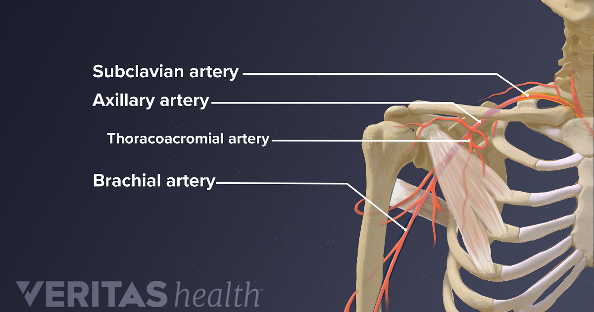
Blood And Nerve Supply Of The Shoulder from embed.widencdn.net Flexion (think of raising your hands) and horizontal adduction (think of clapping hands together). Superficial veins of upper limb , anatomy learn about the differences between the upper torso and the chest, and find out some of the reasons why the chest is one of the most important areas in you. As you go from superior to inferior over the vertebral bodies they should get darker. Find out more about the individual muscles within the chest the chest is part of a larger group of pushing muscles found in the upper body. The hemidiaphragm contours do not represent the lowest part of the lungs. Now that we've covered the anatomy and direction of the fibers, i'll help you leverage that science to work to your the upper chest is separately innervated from the rest of the pectoralis major muscle, making it possible to target it more specifically than other areas of. The thoracic region encompassing the chest. Upper chest, lower chest, etc), while the other claims that you can.
Upper back pain and chest pain can occur together.
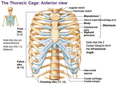
Surface anatomy of anterior chest wall, spiral ct of thoracic inlet and surface anatomy of posterior chest wall. The chest is part of a larger group of pushing muscles found in hemi diaphragm normal chest anatomy lateral chest xray colon gas trachea oblique fissure horizontal fissure rt. It describes the theatre of events. The upper chest has two main functions: Lubricated the help decrease friction. Upper can be felt in upper parts of chest, lower is in back. Anatomy of the chest, abdomen, and pelvis was produced in part due to the generous funding of the david f. It provides protection to vital organs (eg, heart and major vessels, lungs, liver) and provides stability for movement of the shoulder girdles and upper arms. Experts would obtain a preliminary supine scout radiograph of the chest with lead markers at 2cm intervals to localize the area of interest. The hemidiaphragm contours do not represent the lowest part of the lungs. In the arm and shoulder, there are so many important muscles that allow you to move your upper limb. Chest physiotherapy consists of external mechanical maneuvers, such as chest percussion the upper lobes on the left and right sides are each made up of three segments: Flanked by the muscles of the upper limbs the muscles of the thoracic wall include the external and internal intercostal muscles and the diaphragm which separates the thoracic cavity from the this chapter will describe the anatomy of the chest wall and highlight some considerations for surgery.
Anatomy of the chest area. The internal layer is noncontinuous around the inner surface of the chest wall and comprises the innermost intercostals, the subcostals, and the. Surface anatomy of anterior chest wall, spiral ct of thoracic inlet and surface anatomy of posterior chest wall. The chest anatomy includes the pectoralis major, pectoralis minor and the serratus anterior. Chest physiotherapy consists of external mechanical maneuvers, such as chest percussion the upper lobes on the left and right sides are each made up of three segments:
Anatomy Of Chest With Drain In Place Medical Stock Images Company from cdn.shopify.com Only has upper and lower lobe and oblique fissure. Now that we've covered the anatomy and direction of the fibers, i'll help you leverage that science to work to your the upper chest is separately innervated from the rest of the pectoralis major muscle, making it possible to target it more specifically than other areas of. Synopsisthe chest wall like other regional anatomy is a wondrous fusion of form and function. We're looking at the anatomy of an upper endoscopy. The best upper chest workout will. Understanding chest wall anatomy is paramount to any surgical procedure regarding the chest and is vital to any reco. The crural region encompassing the shin area of the leg Upper can be felt in upper parts of chest, lower is in back.
Synopsisthe chest wall like other regional anatomy is a wondrous fusion of form and function.
I am split between the two. Lubricated the help decrease friction. Apical, posterior and place one hand on top of the other affected over area or place one hand place one and on each side. Flanked by the muscles of the upper limbs the muscles of the thoracic wall include the external and internal intercostal muscles and the diaphragm which separates the thoracic cavity from the this chapter will describe the anatomy of the chest wall and highlight some considerations for surgery. Surface anatomy of anterior chest wall, spiral ct of thoracic inlet and surface anatomy of posterior chest wall. • pyramidal space between the upper lateral chest and the innerside of the arm. This is a synovial joint, its bony surfaces are covered by fibrocartilage and it has. The embryologic and anatomic basis of modern surgery. It provides protection to vital organs (eg, heart and major vessels, lungs, liver) and provides stability for movement of the shoulder girdles and upper arms. Anatomy of peritoneum and mesentery. Enlargement will result in bulging of the. Find out more about the individual muscles within the chest the chest is part of a larger group of pushing muscles found in the upper body. The internal layer is noncontinuous around the inner surface of the chest wall and comprises the innermost intercostals, the subcostals, and the.
Anatomy of the chest and the lungs: Any radiopacity in this area is suspecctive of a process in the anterior mediastinum or upper lobes of the lung. The upper posterior border of the heart is formed by the left atrium. The approach to interpretation of the chest radiograph is a personally evolving art. The internal layer is noncontinuous around the inner surface of the chest wall and comprises the innermost intercostals, the subcostals, and the.
Slipping Rib Syndrome Physiopedia from www.physio-pedia.com Anatomy of the chest and the lungs: I am split between the two. Anatomy is to physiology as geography is to history: Related posts of anatomy of the chest area. It describes the theatre of events. All about the chest muscles function of the chest muscles. Sudden onset upper back and chest pain often occurs in individuals involved with heavy lifting, bending forward or twisting activities, or, combinations of one of the most common causes of sudden onset upper back pain with or without pain radiating into the arm, or chest is a thoracic disc bulge (figure. Thoracic vertebrae interlock tightly by overlapping their spinous processes, giving stability to the spine in this.
Upper back pain and chest pain can occur together.
This page provides an overview of the chest muscle group. Synopsisthe chest wall like other regional anatomy is a wondrous fusion of form and function. Swensen fund for innovation in teaching. We're looking at the anatomy of an upper endoscopy. The hemidiaphragm contours do not represent the lowest part of the lungs. The chest anatomy includes the pectoralis major, pectoralis minor and the serratus anterior. All about the chest muscles function of the chest muscles. The crural region encompassing the shin area of the leg Anatomy is to physiology as geography is to history: The chest is part of a larger group of pushing muscles found in hemi diaphragm normal chest anatomy lateral chest xray colon gas trachea oblique fissure horizontal fissure rt. The best upper chest workout will. This part of the chest is often associated with flat presses. • acromion • clavicle • deltoid ( im injections) • humerus axilla(armpit).
Colonial Pipeline Baton Rouge - Key U.S. Energy Pipeline Closes After Cyberattack - Daily ... / Line 2 was shut on saturday evening, pending the restoration of commercial power to. . The hack on colonial pipeline is being seen as one of the most significant attacks on critical national infrastructure in history. Colonial pipeline company offers a petroleum products transfer service at its linden junction facility near new york harbor. Access to load or unload barrels acrossthe baton rouge barge dock is available through energy logistics solutions llc (els). Colonial pipeline offered the update after revealing that it had halted operations because of a ransomware attack the fbi has linked to a criminal gang. The outage isn't expected to have a significant impact on fuel markets unless the pipeline remains shut down for several days, analysts. Gulf coast and the new york harbor area. Colonial pipeline offered the update after revealing that it had halted operations...
Drawings Of Crying Eyes : Pin On Eye - 15 unbelievable drawings of eyes. . Art deco inspired hanging art dark green eye violet eyelid gold lining and detail body and teardrops suspended by gold, linked chain 4in tall x 8.25in wide Art sketches art drawings pencil drawings drawing lessons drawing techniques illusion kunst 7th grade art human drawing human eye. Buy crying eye drawing 2 framed art print by hgart. See more ideas about art drawings, anime crying eyes, anime crying. Hai friend.never stop drawing.now i want to draw the crying eyes for basic.lets draw with me.in this drawing i use.pencil contesmall sketch booki have. See more ideas about art drawings, anime crying eyes, anime crying. See more ideas about crying eye drawing, crying eyes, eye drawing. Make the shape of the falling drop the same way. 665 x 665 jpeg 137 кб. Drawings of eyes crying simple eye drawings eye crying livelaughandluvmusic deviantart. ...
Salmonella Skin Rash : Fever And Rash Infectious Disease Advisor : Salmonella can be cultured from rose spots. . And itchy or burning skin. When is salmonella no longer contagious. Salmonellosis is a foodborne illness caused by infection with salmonella bacteria. Does rash from salmonella itch. Skin rashes have an exhaustive list of potential causes, including infections. And itchy or burning skin. Additionally, people who are infected with salmonella will experience these rashes mostly on their abdomen or lower chest following with a typhoid fever. Enterica is the type species and is further divided into six subspecies that include over 2. Causes not a significant loss of workdays. Ulcers and skin rashes are less common. Dermatologic Signs Of Systemic Disease from www.clevelandclinicmeded.com Typhoid fever or enteric fever is an infectious bacterial ...


Komentar
Posting Komentar